14. INTERNATIONALE MEDIZINISCHE C-SCHUTZ-TAGUNG 2013 (POSTERABSTRACTS, TEIL2)
WMM, 57. Jahrgang (Ausgabe 8-9/2013: S. 224-231)
From the laboratory to the field: cholinesterase phosphylation and its subsequent reactivation by oximes in correlation with temperature*
Katalinić M¹, Šinko G¹, Kovarik Z¹, Stojan J²
¹Institute for Medical Research and Occupational Health, Ksaverska cesta 2, HR-10000 Zagreb,
Croatia; ²Institute of Biochemistry, Medical Faculty, University of Ljubljana, Vrazov trg 2, SI-1000 Ljubljana, Slovenia
The importance of cholinesterases (AChE, EC 3.1.1.7 and BChE, EC 3.1.1.8) in maintaining the homeostasis of an organism has been investigated and presented ever since their discovery. Even now, many new roles for these enzymes are being considered and described.
Since many of the in vitro experiments concerning cholinesterases are performed at a lower temperature than the physiological one, a translation of the obtained experimental results to in vivo conditions presents a great challenge. Namely, activity of cholinesterases as enzymes displays a certain temperature optimum, in other words, all kinetic parameters describing cholinesterase activity and their interactions with ligands are temperature dependent. We focused our study on the thermodynamic characterization of cholinesterase interactions with organophosphorus pesticides and nerve agents, and the subsequent reactivation of phosphylated cholinesterases by oximes. The threat of a misuse of these highly toxic organophosphorus compounds (OPs) is still widely present. On the other hand, oximes as antidotes are characterised as orphan drugs, which further raises a wide array of concerns that should be addressed even in the early phases of antidote development. Therefore, a critical view is required for in vitro obtained experimental results in this area of research. The main goal of our study was to define a temperature-correlated model of cholinesterase phosphylation as well as of oxime-assisted reactivation. Such a model could prove valuable for estimating kinetic data at a desired temperature, for example the physiological one, based on the experiments performed at more convenient temperatures. It would also ensure a better selection of compounds for in vivo experiments. Furthermore, knowing the manner in which the properties of cholinesterases alter within a temperature range could improve the development of cholinesterase-based agents for OP decontamination.
* Acknowledgment: This work was supported by the Croatian-Slovenian bilateral project 2012-2013 (PIs:, G. Šinko,, J. Stojan).
Hospital based decontamination system established in military medical academy for chemical victim
Kenar L, Ortatatli, M, Sezigen S, Kunak ZI
Department of Medical CBRN Defense, Gulhane Military Medical Academy, Ankara, Turkey
CBRN warfare agents are commonly supposed as a global threat which all health care organizations should be prepared for. As one of the measures, hospitals are required to have the capability of performing patient decontamination. The purpose of decontamination unit is to provide the removal of CBRN contamination from the casualties before they enter the clean medical treatment area where maximal medical care can be provided. From this perspective, a permanent medical decontamination unit based on scientific facts and our knowledge and experiences was established in our Military Medical Academy which is the highest level health-care foundation in our country. Decontamination unit is known as an essential structure to clean the patients / victims before accepting them into the interior part of the hospital to furtherly perform advanced and intensive medical care including curative and emergency intervention. The unit is placed adjacent to the Emergency Department and separated into two corridors in which each has sliding stretcher system for unconscious litter victims. The unit is furnished with sufficient amounts of showerheads, water supplies, decon solutions, waste drainage tract to efficiently decontaminate 420 ambulatory patients and 60 unconscious patients who definitely need more time and staff. Since decon of casualties is an enormous work requiring devotion of large numbers of medical personnel and paramedics and a large amounts of time with patience, a total of 13 staff is assigned for medical decontamination. In the entry control point, main function is to perform triage and detection of the patients and the agent along with the prompt resuscitation before allowing patients through decon corridors. In the designated triage part of the unit, physicians assigned will quickly evaluate each casualty and fix them into one of the triage categories.
In this submission, we would like to give more info about this decontamination unit which is a proud of our Academy and nominated as a role model for other military and civilian hospitals.
Anticholinergic properties of MB327 and oximes*
Königer C, Worek F, Thiermann H, Wille T
Bundeswehr Institute of Pharmacology and Toxicology, Munich, Germany
Poisoning by organophosphorus compounds (OP) leads to inhibition of the enzyme acetylcholinesterase (AChE) resulting in impaired cholinergic signalling and overstimulation of cholinergic receptors. At present, standard therapy of OP poisoning includes an oxime as reactivator of inhibited AChE and atropine as competitive muscarinic antagonist. Recent investigations showed that the bispyridinium non-oxime MB327 improves soman-blocked neuromuscular transmission in human intercostal muscle and rodent diaphragm preparations in vitro. Furthermore, MB327 showed a high affinity to muscarinic acetylcholine receptors. Up to now, data on the effects of MB327 in tissue are scarce. Thus, we investigated the anticholinergic properties of MB327 and oximes in a rat jejunum smooth muscle model and compared the EC50s calculated from the respective dose-response curves.
All tested compounds exhibited a concentration-dependent, reversible smooth muscle relaxing effect. Among the pyridinium compounds tested, MB327 showed to be most potent (EC50 = 6 µM). The structural analogue TMB-4 (EC50 = 41 µM), bearing two oxime instead of tert-butyl groups, was the most potent oxime tested followed by obidoxime (EC50 = 90 µM). 2-PAM (EC50 = 249 µM), MMB-4 (EC50 = 264 µM) and HI-6 (EC50 = 711 µM) showed a substantially lower smooth muscle relaxing effect. However, relaxation by atropine (EC50 = 0.015 µM) was much more effective than MB327. Simultaneous administration of 25 nM atropine (resembling a therapeutic plasma concentration) and MB327 (1 – 45 µM) resulted in a pronounced antimuscarinic effect suggesting an additive effect of atropine and MB327. MB327 exhibited an antimuscarinic smooth muscle relaxing effect which could contribute to its therapeutic effect in OP poisoning. In comparison to clinically used oximes MB327 had a 7- to 115-fold higher antimuscarinic potency. At present, the relevance of the antimuscarinic properties of MB327 for its overall therapeutic effect in OP poisoning is difficult to assess. Additional studies are needed to evaluate the potential of MB327 as a broad-spectrum OP antidote and to investigate in detail its mechanisms of action.
* This work has been accepted for publication in Chemico-Biological Interactions on April 9, 2013.
Novel mechanisms in the pathogenesis of late Mustard toxicity
Korkmaz A1, Kunak ZI2
1Gulhane Military Medical Academy, Department of Physiology, Ankara, Turkey; 2Gulhane Military Medical Academy, Department of Medical CBRN Defence, Ankara Turkey
Sulfur Mustard (SM), also known as mustard gas, has been most widely used chemical weapon. The toxicity of SM as an incapacitating agent is much greater importance than its ability to cause lethality. After several decades of studies, the acute toxicity of SM has been revealed as nitro-oxidative stress, DNA alkylation, energy depletion and a consequent robust inflammation with in the affected tissues. Therefore, several antioxidant compounds and anti-inflammatory drugs have been shown beneficial effects on acute toxicity. The delayed toxicity of SM however, has still no mechanistic explanation. Victims of Iran-Iraq war (1981-1989) have been still suffering from late toxic effects of SM exposure including ophthalmic, cutaneous and respiratory sequels. Among them, respiratory disorders are the most frequent and disabling consequences. In spite of the vigorous treatment modalities, physicians have failed to treat late toxic effects of SM exposure although it mimics chronic obstructive pulmonary disease (COPD). Because of the abovementioned reason (i.e., failure to treat with antioxidants and/or anti-inflammatory drugs including both steroids and NSAID) SM-induced lung toxicity has been coined as „mustard lung“. The inability to treat chronic toxicity of SM with conventional treatment modalities let us to reconsider about the pathophysiological mechanism of this chemical weapon. According to our experimental results SM seems to be harmful not only for protein coding genes, but also regulatory part of the DNA, therefore is considered as an „epigenetic disruptor“. The word „epigenetic“ explains reversible heritable changes in gene expression that occurred without alteration in the DNA sequence, but changes that were sufficiently powerful to regulate the dynamics of gene expression. The traditional view that gene and environment interactions control disease susceptibility can therefore, be expanded to include epigenetic reprogramming as a key determinant of origins of several human disease. The „mustard lung“ seems presumably as a consequence of epigenetically disrupted cells following SM exposure. By this way, an epigenetic look at the affected tissues may be a novel hope in the treatment of late toxicity of such an enigmatic chemical weapon.
An analysis of poisoning in children below 14 years of age on the basis of files from the National Poison Information Centre in Lodz, Poland with special focus on trends in pesticides
Krakowiak A1, Piekarska-Wijatkowska A2, Malgorzata Kotwica M2
1Toxicology Unit; Department of Occupational Diseases and Toxicology, Nofer Institute of Occupational Medicine, Lodz, Poland; 2National Poison Information Centre, Nofer Institute of Occupational Medicine, Lodz, Poland
Introduction: Poisonings are responsible for significant part of hospital admissions and therefore, but also due to associated mortality and their high prevalence in the population, they are considered as a major health problem. Acute poisonings in children pose real challenge in the clinical toxicology. The aim of the presented study was to characterize the trends in epidemiology of acute poisonings, particularly in aspect of intoxications with pesticides among children below 14 years of age on the ground of material from National Poison Information Center in Lodz (NPIC), Poland.
Material and methods: this work is based on the statistical data gathered by staff of NPIC during 2001–2010.
Results: most children were poisoned with medications, pesticides and corrosives in analysed period of time. Most poisonings due to medications were by analgesic, antipyretic and antirheumatic drugs. Corrosives and solvents were the most common household products responsible for poisoning in children under 14 years. Poisonings with coumarin derivatives were the most common cause of hospitalization among pesticides. Conclusions: intoxications with pesticides still pose problems in developed countries due to diagnostic and therapeutic difficulties.
Centrally active AChE inhibitors as potential prophylaxis against nerve agents*
Korabecny J1,2, Hamuľakova S3, Babkova K1, Horova A1, Musilek K1,2,4, Spilovska K1, Karasova-Zdarova J1, Daniel J,1,2 Kuca K1,2,4
1Faculty of Military Health Sciences, University of Defence, Hradec Kralove, Czech Republic, 2Biomedical Research Center, University Hospital of Hradec Kralove, Hradec Kralove, Czech Republic, 3Department of Organic Chemistry, Institute of Chemistry, Faculty of Science, P.J. Safarik University, Kosice, Slovak Republic, 4Department of Chemistry, Faculty of Science, University of Hradec Kralove, Hradec Kralove, Czech Republic
Inhibition of AChE by reversibly acting AChE inhibitors (AChEIs) is one of the therapeutic approaches in prophylaxis against nerve agents. Within our contribution we are presenting synthesis, biological evaluation (AChE and BuChE inhibition) and molecular modeling of the novel series of tacrine-7-methoxytacrine heterodimers as possible centrally active prophylactics. As resulted several candidates reached promising results comparable with those obtained for standard tacrine. Selected chemical structures will be in the future leading structures for in vivo prophylaxis evaluation.
*Acknowledgement: This work was supported by a long-term organization development plan 1011.
Two promising molecules, melatonin and S-Methylisothiourea as prophylaxis against nitrogen mustard induced intestine toxicity
Kunak ZI1, Yaren H1, Yaman H2, Macit E3, Ongoru O4, Uysal B5, Akgul EO2, Korkmaz A5, Kenar L1
1Gulhane Military Medical Academy, Dept. of Medical CBRN Defense, 2Gulhane Military Medical Academy, Dept. of Medical and Clinical Biochemistry, 3Gulhane Military Medical Academy, Dept. of Analytical Toxicology, 4Gulhane Military Medical Academy, Dept. of Pathology and 5Gulhane Military Medical Academy, Dept. of Physiology, Ankara, Turkey
Objective: Sulfur mustard known as mustard gas is still a great threat to humanity as there is no specific treatment of cure for it yet. Although many studies are being made for improvements, studying with nitrogen mustard is safer and legally easier. One of our studies showed glimpses of hope, but the structure of the experiment demanded on after exposure treatment. Nitric oxide and peroxynitrite are responsible for mustard induced toxicity. In our previous study, it was aimed to compare the therapeutic efficiencies of melatonin (wide-spectrum antioxidant) and S-methylisothiourea (selective iNOS inhibitor) in intestine toxicity caused by nitrogen mustard in rats.
Material and method: Thirty six rats were randomly divided into four groups: control, MEC, MEC+MEL, and MEC+SMT. Group 1 was given 2 ml saline, other groups received single dose of mechlorethamine (MEC) (3.5 mg/kg transdermally) with the same time intervals. Group 3 and 4 received MEL (100 mg/kg i.p.) and SMT (50 mg/kg i.p.), respectively. In all groups, tumor necrosis factor-α (TNF-a), interleukin 1-beta (IL-1b) and nitrate/nitrite (NOx) levels were measured in intestine tissues.
Results: Mechlorethamine injection resulted in severe inflammatory cell infiltration, ischemic changes and vacuolization in small intestine. In melatonin group, vacuolization was decreased, inflammation was minimal in intestine. In S-methylisothioure group, Epithelial damage was minimal and no inflammation in intestine. TNF-a, IL-1b and NOx levels in the MEC and MEC+SMT groups were significantly higher compared to the other groups. In terms of TNF-a and NOx concentrations, there was no difference between the control and MEL groups. There was no difference in the MEC and MEC+SMT groups in terms of IL-1b levels. IL-1b concentrations were found lower in MEL group compared to MEC and MEC+SMT groups. But unfortunately IL-1b levels in MEL group were higher than control group. Increased TNF-a, IL-1b and NOx levels were in good agreement with histopathological injury.
Conclusion: These results suggest that MEL treatment found beneficial. But the prophylactic effects of these two molecules are vague and worth to investigate with further studies.
Structural analogs of the HI-6 oxime are the most potent reactivators of the soman-inhibited AChE mutant that resists ageing*
Maček N1, Radić Z2, Taylor P2, Kuča K3, Kovarik Z1
1Institute for Medical Research and Occupational Health, HR-10000 Zagreb, Croatia; 2Skaggs School of Pharmacy and Pharmaceutical Sciences, University of California at San Diego, La Jolla, CA 92093-0650, USA; 3Center of Advanced Studies, Faculty of Military Health Sciences, 500 01 Hradec Králové, Czech Republic
The toxicity of organophosphorus compounds (OPs) caused by the phosphylation of AChE active center serine results in the accumulation of the neurotransmitter acetylcholine (ACh), consequently causing seizures and in severe cases even death. Immediate therapeutic treatment usually consists of a combined administration of anticholinergic drugs and an oxime as the reactivator of AChE. The nerve agent soman is an extremely toxic OP and acts almost instantaneously. AChE inhibited by soman is less accessible to reactivation due to the rapid dealkylation phenomenon of the soman-AChE conjugate called ˝ageing˝. Earlier studies have shown that amino acid substitutions in the AChE active center could slow down ageing and enable oximes to reactivate the soman-AChE conjugate prior to its ageing. The human AChE mutant hY337A/F338A combines the increase in the active center accessibility of the Y337A mutant to oximes with the ageing resistance of the F338A mutation. Therefore, its administration to potentially exposed individuals in the form of a pre-treatment is suggested so that it acts as a pseudo catalytic bioscavenger. Nevertheless, apart from ageing, the oxime reactivation of the soman-AChE conjugate is effected by the steric hindrance of the soman’s alkoxy side chain which limits the effectiveness of pyridinium oximes. In the 1970s, it was found that potential reactivators have an oxime group in the ortho position on the pyridinium ring and/or a CH2-O-CH2 linking chain between two pyridinium rings. Oxime HI-6, containing both characteristics, especially stood out. The search for novel oxime reactivators is still ongoing. We tested a number of novel and some previously known oximes in order to challenge the 1970s findings. Nevertheless, among all the tested oximes, HI-6 and its structural analogs, when taking kobs as the main criterion, have proven to be the most potent reactivators of the soman-Y337A/F338A conjugate.
* Acknowledgments: This work was supported by the NIH (Grant No. U01 NS058046) and by the Croatian Ministry of Science, Education and Sports (Grant No.022-0222148-2889).
Monooxime bisquaternary cholinesterase reactivators with 3-oxapent-1,5-diyl linker – preparation and in vitro screening study
Musilek K1-3; Kasparek A1; Horova A1; Herman D1; Krenkova Z1; Jun D1,3; Kuca K1,3
1University of Defence, Faculty of Military Health Sciences, Department of Toxicology and Centre of Advanced Studies, Trebesska 1575, 500 01 Hradec Kralove, Czech Republic; 2University of Hradec Kralove, Faculty of Science, Department of Chemistry, Rokitanskeho 62, 50003 Hradec Kralove, Czech Republic; 3University Hospital, Sokolska 581, 500 05 Hradec Kralove, Czech Republic
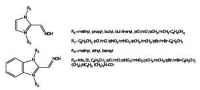
The reactivators of cholinesterases (AChE, EC 3.1.1.7; BChE EC 3.1.1.8) are crucial components in the treatment of intoxications caused by organophosphates (OP) such as nerve agents and pesticides [1]. These OP inhibitors covalently bind to active site of mentioned enzyme and irreversibly inhibit its activity. The reactivator breaks the inhibitor-enzyme covalent bond and restores its activity. Unfortunately, there is no reactivator applicable for every type of OP inhibitor [2]. Formerly, bisquaternary mono-oxime AChE reactivators were recommended against organophosphate poisoning [3]. The series of bisquaternary molecules was prepared (Fig. 1) and evaluated on human erythrocyte AChE or human plasmatic BChE inhibited by different OP (dichlorvos, paraoxon, sarin, VX) in vitro. The method set up excluded the excess of OP within the measurement. The results showed that some prepared compounds were able to effectively counteract OP inhibition in vitro at both tested concentrations (100 and 10 μM).
References: Bajgar J: Adv. Clin Chem 2004, 38, 151. Marrs TC: Pharmacol Therapeut 1993, 58, 51. Musilek K, et al.: Med Res Rev 2011, 31, 548.
*Acknowledgement: The work was supported by the Ministry of Defence of the Czech Republic (a long-term organization development plan 1011).
Novel Antidotal Mixtures against Organophosphate Nerve Agents
Nili U, Katalan S, Shlomi L, Yacov G, Raveh L, Amitai G, Bloch-Shilderman E
Dept. of Pharmacology, Israel Institute for Biological Research, Ness-Ziona, Israel
Nerve agents such as Sarin, Soman and VX are organophosphates (OP) whose toxicity is induced by inhibition of synaptic acetylcholinesterase (AChE). This irreversible inhibition prevents hydrolysis of acetylcholine, leading to its accumulation in synaptic clefts and consequent excessive peripheral and central cholinergic stimulation. The excessive cholinergic activity triggers a rapid progression of toxic effects. These include hyper secretions, tremor, seizure activity and concomitant motor convulsions, which may rapidly progress to status epilepticus and profound brain damage, inhibition of the medullary respiratory center and ultimately death. The current antidotal auto-injector against OP nerve agent intoxication used in Israel consists of the oxime TMB-4 intended to reactivate the inhibited enzyme, and the anticholinergics atropine and benactyzine intended to counter the excessive peripheral and central cholinergic activity, respectively (TAB). The aim of the present work was to develop an improved antidotal therapy against lethality and neuropathology induced by OP nerve agents. A series of in-vivo studies pointed to caramiphen, a drug with a broad pharmacological profile, as superior to other centrally active anticholinecholinergics in protecting against nerve agent induced neuropathology and behavioral deficits. A series of in-vitro studies indicated that HI-6 and MMB-4 are superior to other oximes in reactivating AChE inhibited by OP nerve agents. Consequently, we compared the efficacy of antidotal therapy following Sarin, Soman and VX intoxication in rats, by antidotal mixtures containing the oximes TMB-4, HI-6 or MMB-4, and the anticholinergics atropine together with either benactyzine or caramiphen. Results demonstrated that a combined treatment of HI-6/MMB-4, atropine and caramiphen is superior to the TAB antidotal mixture against OP nerve agent intoxication.
Combination Therapy for Percutaneous Nerve Agent Poisoning*
Price M, Armstrong S, Tattersall J, Mumford H
Biomedical Sciences Department, Dstl Porton Down, Salisbury, Wiltshire, SP4 0JQ, UK
Organophosphate poisoning by the percutaneous (p.c.) route remains a challenge for immediate self- or buddy-aid. We have previously shown, using a guinea-pig model of p.c. poisoning, that stoichiometric bioscavenger (human plasma-derived butyrylcholinesterase, huBuChE) provided effective protection when administered before signs of poisoning appeared. Administration could be delayed until there are signs of systemic poisoning following p.c. nerve agent exposure, if the bioscavenger was supported by a single administration of conventional therapy drugs. In these studies, the dose of huBuChE was limited by practical considerations (the volume that could be administered by the intramuscular (i.m.) route and the protein concentration of the solution). The aim of the present study was to determine the extent to which the degree of protection was dependent on the plasma concentration of bioscavenger, by using intravenous (i.v.) huBuChE dosing as an experimental tool to increase the amount of bioscavenger available in the blood at an early stage following administration (akin to increasing the i.m. dose). It was hypothesised that this might improve the protection ratio of the combination therapy by increasing the amount of HuBuChE available to bind absorbed VX.
Male Dunkin-Hartley Guinea-pigs (Harlan UK) were exposed to a p.c. challenge of VX diluted in isopropyl alcohol, and therapy was administered on the appearance of a specific set of systemic signs of cholinergic poisoning (incapacitation, tremor, salivation, lachrymation). Therapy comprised a single i.m. injection of HI-6 dimethanesulphonate (27.9 mg/kg), atropine sulphate (17.4 mg/kg) and Avizafone hydrochloride (3.14 mg/kg), supported by huBuChE (14980 U/kg) given either i.m. or i.v. An adaptive dosing design was used to determine a protection ratio (PR) for each treatment regimen. The PR (LD50 with MedCM/LD50 without MedCM) of Combination Therapy including i.m. huBuChE was 2.24, and the new results will be discussed in the context of the pharmacokinetic profiles of atropine, diazepam, HI-6 and huBuChE, which are presented in an accompanying poster (Armstrong, 2013).
* © Crown Copyright. Dstl 2013.
Sulfur Mustard Detector – a fast and reliable test kit for on-site detection of sulfur mustard contamination on skin surface*
Ringlstetter S1, Kolb M1, Steinritz D2, Kehe K2@und Klaus S1
1Securetec Detektions-Systeme AG, Eugen-Sänger Ring 1, 85649 Brunnthal / Munich, Germany; 2Bundeswehr Institute of Pharmacology and Toxicology, Munich, Germany
On-site trace detection of warfare agents has become increasingly important for German Armed Forces involved in international military operations such as those in Afghanistan and, most recently, Turkey. The threat of sulfur mustard attacks is most likely presented by those countries which possess bulk quantities of this devastating agent (e.g. in the Middle and Near East) while at the same time, are exposed to known terrorist activities. In order to protect soldiers properly, rapid detection devices are of great need. As part of the strategy to fulfill this need, a Sulfur Mustard Detector device was developed that allows reliable and rapid detection of small quantities of sulfur mustard on skin surfaces. The Sulfur Mustard Detector device is based on an immunochromatographic detection technology that has significant advantages over other detection methods. Most notable is its easy on-site use, its small size of the detector and its rapid test results (in few minutes). A highly sensitive and specific detection of sulfur mustard was mainly achieved by using a unique antibody. Furthermore, the Sulfur Mustard Detector device was developed and optimized to meet all special needs of armed forces demanded in harsh field conditions. The device is small (13.5 x 2.5 x 2 cm), it can be used in temperatures from 5°C to 45°C, the test procedure is quick and easy to handle and the shelf life is 18 months. The lower detection limit is 2 µM sulfur mustard under lab conditions. When testing skin from pigs, the lowest detection amount was 3 pg of free sulfur mustard.
Substantial field tests were done during two NATO exercises in Canada where contaminated living pigs as well as other non-animal surfaces were tested. The results from these filed tests confirmed the reliability and good sensitivity of the device. Especially the easy handling, the fast and reliable detection of sulfur mustard combined with high sensitivity was assessed positively – Even under challenging temperature and environmental conditions. Following successful validation, the Sulfur Mustard Detector is labeled with CE and IVD.
* Acknowledgement: The development of the Sulfur Mustard Detector was funded by the German Ministry of Defense, represented by the Bundesamt für Wehrtechnik und Beschaffung (BWB), contract numbers E/UR3G/5G090/5A804 and E/UR3G/8G083/8A802. Contact: [email protected]
Binding Studies of Organophosphonate Cyclodextrin Complexes
Schneider C, Kubik S
Fachbereich Chemie-Organische Chemie, Technische Universität Kaiserslautern, Erwin-Schrödinger-Straße, 67663 Kaiserslautern, Germany
The biological activity of certain organophosph(on)ates (OPs) was discovered in the 1930s by Gerhard Schrader. The high toxicity of these compounds, examples of which are paraoxon, sarin, tabun, and VX, allows their use as insecticides but also as chemical warfare agents. Current therapies of OP poisonings consist in administration of atropine as an antagonist for acetylcholine and oximes for reactivation of inhibited acetylcholine-esterase.[1] The use of atropine, which is also a nerve agent, clearly shows that an effective and safe treatment of OP poisonings is still lacking.
A promising approach involves the use of scavengers that can cleave OPs before they react with acetylcholinesterase. The work of Estour showed, for example, that modified cyclodextrins, bearing a nucleophilic group as a substituent, are suitable agents to detoxify OPs.[2] Cyclodextrins are cyclic oligosaccharides that possess a hydrophobic cavity allowing them to bind non polar compounds in aqueous solutions. Encouraged by these results systematic work was performed in the Kubik group to develop new cyclodextrin-based scavengers. These investigations showed that β-cyclodextrins containing α-effect nucleophiles along the ring can efficiently cleave cyclosarin or tabun.[3][4]
Degradation of the extremely toxic VX is significantly slower than that of other OPs, however possibly due to the fact that the VX molecule binds only weakly to cyclodextrins. The aim of this work is to synthesize cyclodextrin derivatives bearing hydrogen bond donors along the ring, which can interact with the P=O-group of the OPs to improve complex stability. The influence of the substituent on cyclodextrin affinity is quantified by using isothermal titration calorimetry. This poster introduces the concept, summarizes the synthetic routes to the different cyclodextrin derivatives and shows some results of the ITC-measurements.
References: [1] N Aurbek, H Thiermann, L Szinicz, P Eyer, F Worek: Toxicology, 2006, 224, 91-99. [2] R Le Provost, T Wille, L Louise, et al. : Org Biomol Chem, 2011, 9, 3026-3032. [3] F Brandhuber, M Zengerle, L Porwol, et al.: Toxicology, 2012, 302, 163-171. [4] M Zengerle, F Brandhuber, C Schneider, et al.: J Org Chem, 2011, 7, 1543-1554.
2-PAM and HI-6 efficacy in soman poisoning in the guinea pig: an integrative numerical–experimental investigation
Seng KY, Yap XH, Yeo TH, Tan YT, Emily C-S, Saw XT, Tan SL, Tang FR, Loke WK
DSO National Laboratories, 27 Medical Drive, Singapore 117510
The primary mechanism of in vivo toxicity for highly toxic organophosphates, such as nerve agents (NA), is the inhibition of acetylcholinesterase (AChE). Reactivation of NA-inhibited AChE, an important step in the treatment of NA poisoning, is the main therapeutic mechanism of oximes such as Pralidoxime (2-PAM) and HI-6. Recently, a numerical model that integrates AChE activity (inhibition, reactivation and aging kinetics) with NA toxicokinetics and oxime pharmacokinetics was used to calculate the proportion of uninhibited AChE following NA challenge [1]. The main objectives of this study were to (1) estimate – through numerical model simulations – single, intramuscular (i.m.) HI-6 dose(s) that could produce at least 10 % uninhibited blood AChE within 30 min of single, supralethal soman (GD) challenge (115 µg/kg, intravenous [i.v.]) in the guinea pig; (2) predict uninhibited AChE kinetics after treatment with equimolar 2-PAM dose(s) under similar GD challenge conditions; and (3) verify the efficacy of model-predicted HI-6 and/or 2-PAM doses for GD challenge in vivo. The numerical model was parameterised using published GD toxicokinetic data [2] and blood AChE rate constants [3]. HI-6 and 2-PAM pharmacokinetic parameters were obtained from in house pharmacokinetic experiments and from [1], respectively. Results from the numerical model showed that a single 250 mg/kg i.m. HI-6 dose administered at t = 1.25min could produce 16.2, 13.7 and 12.2 % uninhibited AChE in the 500, 600 and 700mg guinea pig, respectively, 30min after the supralethal GD challenge. In contrast, the equimolar single i. m. dose of 2-PAM (114 mg/kg) was predicted by the numerical model to produce < 10 % uninhibited AChE across all evaluated guinea pig models. The efficacy of the 250 mg/kg HI-6 dose was subsequently validated based on guinea pig survival data (n = 2). In addition, our in vivo experiments also verified that no guinea pig survived after treatment with either 75 mg/kg HI-6 or 114 mg/kg 2-PAM following GD exposure. Based on these analyses, it is surmised that the numerical-experimental modelling approach is promising as a “learn–and–confirm” tool to design and optimise oxime doses for treatment against NA exposure.
To further test and extend the model, additional work could be conducted to evaluate and verify the accuracy of oxime dose predictions in more animals and/or in a large animal model, e.g. swine.
References: [1] Worek et al (2005). Toxicol Appl Pharmacol 209:193-202. [2] Benschop and De Jong (1991). Neurosci Biobehav Re. 15:73-77. [3] Luo et al (2007). Biochemistry 46:1177
Organophosphate Poisonings in View of the Poison Information Centre Freiburg (VIZ)
Stedtler U, Hermanns-Clausen M
Poisons Information Centre Freiburg, University Hospital Freiburg, Germany
Objective: Is there a change in the frequency of calls to the VIZ concerning exposures to organophosphates? Is there a change in the severity of these cases?
Method: Retrospective analysis of the VIZ database covering the years 2000 to 2012. Results: 549 exposures to organophosphates were reported to VIZ during 2000 to 2012. The share of exposures to organo - phos phates among all reported poisoning cases steadily declined, from 5.3/1000 in 2000 to 1.1/1000 in 2009. It increased slightly since then to 1.7/1000 in 2012. The severity of the reported symptoms according to the ‘Poisoning Severity Score’ was graded moderate or severe in 61 cases. Nine of these patients died. The proportion of moderate or severe poisonings to all cases due to organophosphates was 11 %. It is nearly twice the proportion of moderate to severe intoxications in all exposures reported to the VIZ, which is 6 %. However, information regarding the course more than 12 hours after exposure was available in 214 cases only. These cases include 48 of the moderate or severe intoxications (79 %). Most of these 48 cases were caused by oxydemeton-methyl (16) parathion (11) or organophosphates without further differentiation (10). The frequency of cases with oxydemeton-methyl decreased since 2005, as did the frequency of parathion cases. The frequency of moderate or severe organophosphate poisonings decreased from 0.36/1000 in 2000 to 0.16/1000 in 2012.
Conclusion: With regard to the VIZ organophosphate poisoning is infrequent. The frequency of these intoxications is decreasing since 2000. In this group the fraction of moderate to severe poisoning is higher than in the VIZ cases in general.
Efficiency of novel heterocyclic oximes in reactivation of paraoxon-inhibited cholinesterases
Morasi Pipercic S, Makaric S, Krešimir Baumann K, Primožic I, Tomic-Pisarovic S
Department of Chemistry, Faculty of Science, University of Zagreb, Horvatovac 102a, HR-10000 Zagreb, Croatia
Toxic cholinesterase inhibiting compounds like organophosphates (OP) and carbamates are intensively used and produced throughout the world and continue to be responsible for poisoning and deaths.1,2 Chemical warfare agents are of outmost concern due to their possible use in wars and terrorist attacks. Since there is still no single, broad-spectrum compound suitable for the antidotal treatment of poisoning with all OP agents a series of novel, aliphatic and aromatic quaternary salts of N-alkyl/aryl imidazol- and benzimidazole-2-aldoximes have been prepared as potential reactivators of inhibited cholinesterases.
N-alkylation of commercially available heterocyclic azoles resulted in their N-alkyl/aryl derivatives. N-alkyl/aryl imidazoles and benzimidazoles were converted to the appropriate 2-carbaldehydes by using modified Ivesen-Lund method3 and then to 2-aldoximes. Reaction of the oximes with the alkyl/aryl halides resulted in a desired quaternary salts. Structures of prepared compounds were deduced from IR, one- and two-dimensional NMR and MS spectra. Elman’s method and acetylthiocholine as substrate was used to test inhibitory properties of prepared compounds and their reactivation efficiency of Paraoxon™ inhibited human acetylcholinesterases and butyrylcholinesterases. Structural characteristics of an efficient oxime reactivator will be discussed.References: [1] Primožic I, Odžak R, Tomic S, Simeon-Rudolf V, Reiner E: J med chem def 2 (2004) 1-30. [2] Odžak R, Calic M, Hrenar T, Primožic I, Kovarik Z: Toxicology 233 (2007) 85-96. [3] Iversen PE, Lund H: Acta Chem Scand 10 (1966) 2646-2657.
Nanoparticles for the transport of oximes over the bloodbrain barrier*
Wagner S1, Dadparvar M2, von Briesen H1 , Kreuter J2
1Fraunhofer Institute for Biomedical Engineering (IBMT), Department of Cell Biology& Applied Virology, Ensheimer Strasse 48, 66386 St. Ingbert, Germany; 2Goethe University, Institute of Pharmaceutical Technology, Max-von- Laue-Str. 9, 60438 Frankfurt/Main, Germany
In the case of organophosphorous nerve agent poisonings an immediate administration of acetylcholinesterase (AChE) reactivating antidotes like obidoxime or HI6 is required. However, these oximes are unable to rapidly penetrate the blood-brain barrier (BBB). The BBB protects the brain from the peripheral circulation and represents an insurmountable obstacle for most drugs. Nanoparticulate bound drugs are a solution for this problem. The objective of this work was to load oximes to human serum albumin (HSA) nanoparticles (NP). These NP have previously been shown to enable the delivery of drugs to the brain that normally cannot penetrate the BBB. Therefore, this research project was to improve the efficiency of oximes by developing nanoparticulate formulations as carriers for the transport over the BBB. HSA-NP were loaded with obidoxime or HI6 by incorporating the drug into the particles during particle preparation as well as by adsorption to the surface of NP [1] [2]. The release of obidoxime and HI6 from the NP and the transport over the BBB could be shown by cell culture experiments [3]. In a transwell experiment, the BBB was simulated by a monolayer of brain capillary endothelial cells. The upper compartment of the transwell represented the blood side whereas the lower compartment represented the brain side. During the experiments, the medium of the lower compartment were collected after NP incubation and used for further drug release studies like the AChE-reactivation assay. In our experiments, we could show a reactivation of poisoned AChE after incubation with this collected [3]. This result indicates a transport of obidoxime and HI6 over the BBB model by the use of NP. For more detailed information concerning the concentration of the transported drug experiments with 14C-labeled HI6 are ongoing and will be presented at the poster. Furthermore we could clarify the underlying transport mechanism over the BBB [4]. Our studies confirmed an active receptor- mediated endocytotic uptake of apolipoprotein E (ApoE)- modified nanoparticles. The involvement of low density lipoprotein receptor family members, notably the low density lipoprotein receptor related protein 1 (LRP1), on the uptake of the ApoE-modified nanoparticles into the brain capillary endothelial cells could be shown.
References: [1] Kufleitner et al.: Adsorption of obidoxime onto human serum albumin nanoparticles: Drug loading, particle size and drug release. J Microencapsulation 2010, 27 (6): 506-513. [2] Dadparvar et al.: HI 6 human serum albumin nanoparticles - development and transport over an in vitro blood-brain barrier model. Toxicology Letters 2011, 206, 60-66. [3] Wagner et al.: Nanoparticulate transport of oximes over an in vitro blood-brain barrier model. PLoS One, 2010, 5(12), e14213. [4] Wagner et al.: Uptake Mechanism of ApoE-Modified Nanoparticles on Brain Capillary Endothelial Cells as a Blood- Brain Barrier Model. PLoS One, 2012, 7(3), e32568.
* Acknowledgement: This project was funded by the German Bundesamt für Wehrtechnik und Beschaffung [Förderkennzeichen U2.3 E/UR3G/5G031/5A802].
Determination of warfare agents (nerve agents, blister agents, and ricin) in food, water and environmental samples
Weber W, Schulz H
Zentrales Institut des Sanitätsdienstes der Bundeswehr München, Außenstelle Munster, Laborgruppe Chemie der Gifte / Kampfstoffanalytik; E-mail: [email protected]
Aims: The nerve agents sarin, soman, cyclosarin and VX, the blister agents mustard gas and lewisite, as well as saxitoxin and ricin are substances listed in Schedule 1 of the Chemical Weapons Convention, a disarmament agreement from the year 1997. Signatory states are not permitted to produce or stockpile such substances; however, they remain a possible threat from terrorists or criminals. Nerve agents have a high acute oral and dermal toxicity as they irreversibly inhibit the action of acetylcholinesterase in the synaptic cleft, thus causing a cholinergic crisis. Mustard gas and lewisite are highly toxic to the skin and mucous membranes including the intestinal mucosa.While ricin can easily be obtained from castor beans even in large quantities, and shows very high pulmonary and oral toxicity, saxitoxin, which is naturally produced by shellfish, is not available in significant quantities and cannot be synthesized. For this reason, the latter is unlikely to pose a risk. To address potential threats methods must be developed to verify a putative use of these warfare agents in the investigation of crimes and terrorist attacks.
Materials and methods: As reference substances for chemical warfare agents certified materials were available. For the quantitative determination of nerve agents the Ellman assay was applied. In brief, electric eel acetylcholinesterase and the alternative substrate acetylthiocholine are mixed. Enzymatic cleavage releases thiocholine, which in turn thiolytically liberates yellow 5-mercaptobenzoic acid from 5,5’-dithiobis-(2-nitrobenzoic acid) (DTNB). Product formation is followed photometrically and present nerve agents would cause inhibition of enzyme activity and thus color development. The presence of mustard gas can again be investigated using a photometric method utilizing the alkylation of p-nitrobenzylpyridine giving a colored product. The arsenic blister agent lewisite is detected based on the color reaction of copper(I) with acetylene, which is released from the agent by alkaline hydrolysis. Ricin can be determined in a semi-quantitative manner using a rapid lateral flow immunological assay.
Results and discussion: The developed methods allow the examination of water samples but also of complex matrices such as food, biological and environmental samples. All methods can be carried out without complex equipment and in a short time. The detection limit of all nerve agents in aqueous samples is 0.1 µg/L, for mustards 20 µg/L, for lewisite 50 µg/L and for ricin 10 µg/L. All procedures are accredited methods according to DIN EN ISO 17025.
Conclusions: Methods were developed for the detection of chemical warfare agents in water, food and environmental samples that can be used in the investigation of crimes and terrorist attacks. The detection limits are significantly below the acute toxic doses.
Keywords: warfare agents; nerve agents; blister agents; ricin
Self-regeneration of neuromuscular transmission following soman poisoning in nerve-muscle cell cultures
Weimer I1,2 Eckle V-S1, Seeger T2, Grasshoff C1, Thiermann H2, Antkowiak B1
1Department of Anaesthesiology and Intensive Care, Experimental Anesthesiology Section, Eberhard-Karls-University, Waldhoernlestraße 22, 72072 Tuebingen, Germany; 2Bundeswehr Institute of Pharmacology and Toxicology, Munich, Germany
Organophosphorous compounds depress neuromuscular transmission by inhibiting acetylcholinesterases. In patients suffering from organophosphorous compound poisoning, antidotal treatment, in order to reactivate the enzyme, may have limited success and de novo synthesis of acetylcholinesterases is required to reestablish neuromuscular transmission. However, recovery may take days to weeks, depending on the severity of intoxication. Unfortunately, specific medical therapies that are capable of supporting self-regenerating processes are not available so far. For preclinical development of such treatments a model system is required that allows (i) quantification of recovery of neuromuscular transmission after intoxication with organophosphorous agents and (ii) well controlled pharmacological manipulation of self-regenerative processes. Here we tested the hypothesis that co-cultures of spinal cord and muscle tissue, prepared from embryonic mice, can be used for this purpose, as they contain motor neurons and muscle cells which represent the basic elements of neuromuscular transmission. During the first two weeks ex vivo, synapses were formed between spinal motor neurons and muscle cells and action potential firing of motor neurons caused detectable muscle contractions. Muscle activity was quantified by video microscopy. The frequency of spontaneous muscle contractions was 0.94 ± 0.15 Hz (mean ± sem, n = 19) before the tissue was exposed to soman (final concentration of the nerve agent: 10-5 mol/L, duration of exposure: 20 minutes). After the exposure, muscle activity dropped to 0.20 ± 0.06 Hz. Antidotes were not provided at any time. Yet, three days after soman intoxication, spontaneous muscle activity displayed significant recovery by regaining a mean frequency of muscle contractions of 0.82 ± 0.18 Hz, which remained stable during the next 11 days. In summary these results suggest that self-regeneration of neuromuscular transmission and muscular function is attainable in nerve-muscle-cultures, creating a convenient tool to research pharmacological treatments addressing the de novo synthesis of acetylcholinesterases.
Organophosphonat Degradation by Cyclodextrins Modified with Hydroxamic Acids
Zengerle M1, Brandhuber F2, Bierwisch A2, Kubik S1
1Technische Universität Kaiserslautern, Fachbereich Chemie-Organische Chemie, Erwin-Schrödinger Straße, 67663 Kaiserslautern, Germany; 2Institut für Pharmakologie und Toxikologie der Bundeswehr, Munich, Germany.
Several organophosph(on)ates (OP) such as sarin, tabun, paraoxon, or VX possess high acute toxicity which is why these compounds are used as chemical warfare nerve agents or pesticides. Toxicity is mainly related to the covalent modification of the enzyme acetylcholinesterase (AChE) which plays a key role in neurotransmitter regulation. Current methods to treat OP poisonings involve administration of a combination of atropine and different oximes, the latter of which cause reactivation of AChE, but these therapies have limitations. An alternative therapy for OP poisonings comprises the use of stoichiometric or catalytic scavengers that rapidly degrade the nerve agents in the body before they react with AChE. In this context, appropriately substituted cyclodextrins were shown to possess promising properties.[1] Cyclodextrins, cyclic oligosaccharides composed of 1,4-a- linked glucose subunits, are an excellent basis for the design of enzyme mimics.[2] Their hydrophobic cavity and hydrophilic outside also renders them a good basis for the development of OP scavengers. The general strategy involves introduction of nucleophilic groups along the cyclodextrin ring that mediate OP decomposition once an OP molecule is included into the cavity. A screening of over 90 cyclodextrin derivatives containing diverse nucleophilic groups showed that a-effect-nucleophiles in particular possess a remarkable activity to degrade OPs.[3, 4] Cyclodextrins modified with certain hydroxamic acids along the lower rim can, for example decompose Tabun with a half time of two minutes. Moreover, preliminary results indicate that these scavengers also degrade the extremely toxic VX, which is hydrolytically very stable. This poster shows our concept, summarizes synthetic and analytical approaches, and presents the most promising results.
References: [1] R Le Provost, T Wille, L Louise, et al.: Org Biomol Chem 2011, 9, 3026-3032. [2] R Breslow, DS Dong: Chem Rev 1998, 98, 1997-2011. [3] M Zengerle, F Brandhuber, C Schneider et al.: Org Chem 2011, 7, 1543-1554. [4] F Brandhuber, M Zengerle, L Porwol, et al.: Toxicology 2012, 302, 163-171.
Datum: 25.10.2013
Quelle: Wehrmedizinische Monatsschrift 2013/8-9


