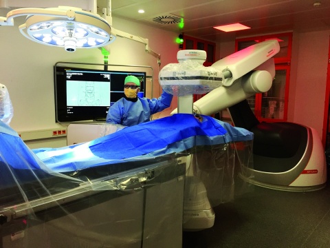
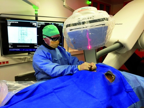
Figure 1: Robot-assisted angiography system
Kai Nestler¹, Benjamin V. Becker¹, Daniel A. Veit¹, Erwin Kollig², Stephan Waldeck¹
The rapid progress of radiological technology and robotics opens up new opportunities for the interdisciplinary treatment of surgical and internal medicine conditions that thus far have been managed by a single area of specialisation.
Two case studies of vertebral body injuries in patients treated at the Bundeswehr Central Hospital in Koblenz are used to demonstrate the opportunities presented by the interdisciplinary use of robot-assisted radiological systems. The authors also discuss how this technology could be used to treat mission-related injuries.
Keywords Percutaneous vertebroplasty, kyphoplasty, needle guidance, Dyna CT, radiofrequency-targeted vertebral augmentation
The rapid progress in radiological technology opens up new opportunities for the interdisciplinary treatment of conditions that were previously managed by a single area of medical specialisation. A good example of this approach is the treatment of spinal conditions such as traumatic fractures and potential vertebral fractures caused by metastatic disease. Such conditions are now being managed by robot-assisted angiography at the Bundeswehr Central Hospital in Koblenz.
Interdisciplinary minimally invasive therapy, e.g. kyphoplasty or spinal fusion using fluoroscopic x-ray guidance, is increasingly being employed by physicians [2]. This is a result of the progress made in this therapeutic approach and the effectiveness of this low-risk treatment and is reflected in the increased number of operations.
Robot-assisted fluoroscopy expands previous treatment options and makes surgery more accurate with fewer complications. It does not involve the restrictions of intraoperative fluoroscopy using a conventional C-arm [8] (Figure 1).
A probe is then used to prepare the vertebral body. The cavities created are filled by way of radiofrequency-targeted vertebral augmentation [4]. In this procedure, radiofrequency is used to activate high-viscosity cement that then hardens. Combined with exact position control, this procedure reduces risks such as extraosseous cement leakage [9].
This highly precise intervention also enables the targeted management of osseous metastases in order to prevent and treat pathological fractures in the spinal and pelvic areas. An interdisciplinary tumour board decides on the treatment, which is then conducted on a multidisciplinary basis. In this context, radiofrequency ablation increases the treatment options available, as a robot can assist with the exact placement of the probe.
We present the cases of two patients who received interdisciplinary treatment with minimally invasive technology and robot-assisted fluoroscopy. One patient had a traumatic vertebral body injury, the other one had painful osseous metastases that threatened the stability of the spine. Neither patient experienced complications. Peri-interventional Dyna CT made it possible to assess the outcome of treatment even before the intervention was completed.
Case 1
Traumatic fracture of the 12th thoracic vertebral body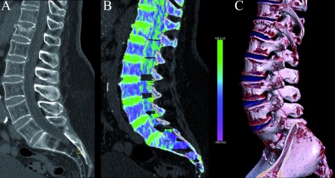 Figure 3:
A: Fracture of the 12th thoracic vertebral body
B: Dual energy CT scans with vertebral oedema
C: 3D visualisation by means of volume rendering
Figure 3:
A: Fracture of the 12th thoracic vertebral body
B: Dual energy CT scans with vertebral oedema
C: 3D visualisation by means of volume rendering
Soldier, aged 37, after a fall
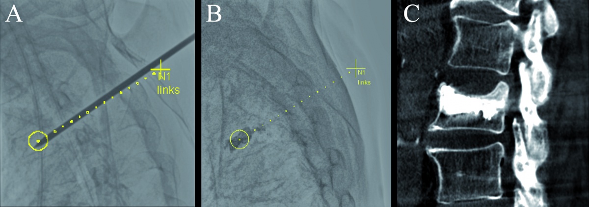
Figure 3A shows an endplate trauma fracture of the anterior edge. A dual energy CT was used to visualise the new vertebral oedema (Figure 3B). A 3D reconstruction (Figure 3C) shows a wedge-shaped vertebral body with a wide base.
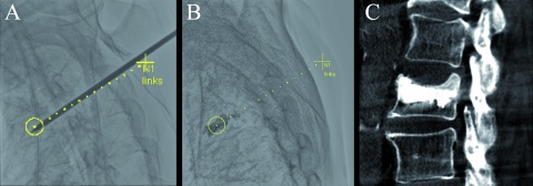
A 3D rotational data set was generated for a final assessment and showed that the vertebral body was sufficiently filled, the endplate was successfully raised, and the position of intravertebral cement was suitable (Figure 4C).
Case 2
Osseous metastasis of the sacrum which may have compromised spinal stability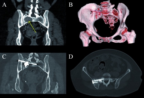 Figure 5:
A: Osteolytic metastasis of the right sacrum (arrow); treatment by means of radiofrequency ablation
B-D: Radiofrequency-targeted vertebral augmentation to fill the affected area and to stabilise the pelvis by screw fixation of the sacroiliac joint
Figure 5:
A: Osteolytic metastasis of the right sacrum (arrow); treatment by means of radiofrequency ablation
B-D: Radiofrequency-targeted vertebral augmentation to fill the affected area and to stabilise the pelvis by screw fixation of the sacroiliac joint
Female patient, aged 58, with osteolytic bone metastases from non-small-cell lung cancer
Figure 5A shows painful osseous metastases on the right side of the sacrum which may have compromised spinal stability. The tumour board decided on multidisciplinary treatment.
To treat the lytic lesion, a probe was first placed using robot-assisted radiofrequency ablation. Radiofrequency-targeted vertebral augmentation was performed via a lateral access route to stabilise the affected area (Figure 5C). The pelvis was stabilised immediately afterwards by screw fixation of the sacroiliac joint via the lateral iliac bone (Figures 5B–D). Stabilisation and a substantial reduction in pain were achieved by the combined therapy.
The treatment of complex conditions demands increased interdisciplinary cooperation, particularly on account of the rapid technical progress made in robot-assisted fluoroscopic systems. The cases presented here show that low-risk operations with good outcomes are possible. Complications, particularly nerve and vascular injuries, and the risk of cement leakage are reduced [1]. The interdisciplinary approach also broadens the range of treatment options and opens up additional opportunities for using osteosynthesis [3]. Especially peri-interventional assessments by means of Dyna CT can lead to new uses in future.
Auxiliary systems such as needle guidance allow surgeons with less practical experience to perform safe surgery. In addition to their timely use in Bundeswehr hospitals after aeromedical evacuation, such systems could also be used in the future to expand treatment options in deployed settings.
Literature
Images: Department of Radiology, Bundeswehr Central Hospital, Koblenz
Citation
Nestler K, Becker BV, Veit DA, Kollig E, Waldeck S: Interdisciplinary Treatment of Spinal Injuries and Bone Metastases Using 3D Robotic Fluoroscopy Wehrmedizinische Monatsschrift 2019; 63(2): 86-88.
For the authors
Dr. Kai Nestler, Squadron Leader (MC)
Bundeswehr Central Hospital, Koblenz
Department of Radiology
Rübenacher Str. 170, 56072 Koblenz
Email: kai1nestler@bundeswehr.org
Datum: 26.04.2019
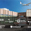
Am Dienstag ist eine automatisiert betriebene Flugzeugdrohne vom Klinikum Ingolstadt aus nach Pfaffenhofen geflogen und hat damit den Grundstein für die Entwicklung neuer Transportwege für…
Die Psoriasis vulgaris ist eine häufige, polygenetische, chronisch-entzündliche Autoimmunerkrankung der Haut und kann durch multiple exogene und endogene Stimuli provoziert werden.
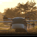
Die Ergebnisse der ersten Missionsflüge bestätigten nicht nur unsere hohen Erwartungen, sondern konnten auch unsere Vision von UAV-basierten Rettungseinsätzen praktisch umsetzen.

Eine Umfrage zeigt, dass beim Thema Wundbehandlung nach wie vor erhöhter Aufklärungsbedarf bei Patienten besteht: Demnach versorgen mehr als zwei Drittel der Befragten ihre Alltagsverletzungen…
Die Vitiligo, häufig auch unter dem Synonym „Weißfleckenkrankheit“ bekannt, ist eine chronische Erkrankung der Haut, bei der es unter Einfluss verschiedener Faktoren zu Funktionsverlust und…
Schifffahrtmedizin an Bord ist durch limitierte diagnostische und therapeutische Ressourcen gekennzeichnet. Daher sind auf See für die gefährdungsminimierende Erstbehandlung sowie…
Nur Maßnahmen, die im Training oft genug wiederholt werden, prägen sich ein und laufen im Ernstfall automatisch ab (SkillDrill). Je realistischer Übungsmöglichkeiten sind, desto einfacher ist der…
Die Behandlung von malignen Tumoren in der Dermatologie hat sich in letzter Zeit stark gewandelt. Die jahrzehntelang einzigen Therapieoptionen mit Chemotherapeutika und Interferon, welche mit einem…
Straumann bietet mit botissCARE Vitamin D eine praktische Chairside Lösung zur Vitamin-D-Wert-Bestimmung sowie ein leicht einzunehmendes und wirksam absorbierbares Vitamin D3+K2 Spray zur Vorbeugung…

Die Hälfte der Weltbevölkerung ist heute dem Risiko ausgesetzt, sich mit dem Dengue-Virus zu infizieren. Überträgerinnen des Virus sind die vorwiegend tagaktiven Mücken der Gattung Aedes aegypti…